
Ultrasound

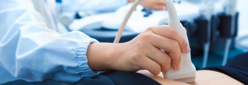
 Our technologist is highly trained in all aspects of ultrasound imaging in order to provide our patients with quality care. The technologist is also available to answer any of your questions and will be with you during the entire examination. An ultrasound, or sonogram, is an image produced by high-frequency (ultrasonic) sound waves to visualize soft tissue structures, organs, and blood vessels within the body. These images can help provide valuable information for diagnosing and treating a variety of diseases and conditions.
Ultrasound imaging requires skin to probe contact. Imaging cannot be done through clothing or material. Afterward the radiologist will interpret the exam and the results will be sent to the ordering physician.
If you have questions about your procedure or cannot keep your appointment date/time, please call our scheduling staff at:
402-460-5884
There are many different types of ultrasound exams and procedures. Listed below are some of the most commonly performed exams:
- Vascular Ultrasounds
- Abdominal Ultrasounds
- Superficial Ultrasounds
- OB/GYN Ultrasounds
- Interventional Procedures
Vascular Exams
Venous Doppler of Arms/Legs
Imaging of the superficial and/or deep veins of the arms and legs to assess for deep vein thrombosis (DVT), also referred to as blood clots, and vascular reflux.
- Duration: these exams can last up to 30 minutes per limb
- Prep: no prep required. All wounds will need to be unwrapped for exam
- Positioning: requires patient to lay flat for duration of exam
Arterial Doppler of Arms/Legs
Imaging of the arteries of the arms and legs to asses for narrowing or blockage of the vessel.
- Duration: these exams can last up to 30 minutes per limb
- Prep: no prep required. All wounds will need to be unwrapped for exam
- Positioning: requires patient to lay flat for duration of exam
Ankle-Brachial Indices (ABI)
Use of blood pressure cuffs placed on arms, ankles and great toes to assess blood flow of the lower extremities. Exam may or may not include a 5 minute interlude of low impact exercise.
- Duration: could last up to 1 hour
- Prep: no prep required
- Positioning: requires patient to lay flat for duration of exam
Carotid
Imaging of the carotid arteries, located on both sides of the neck, to assess for narrowing or blockage of the vessel.
- Duration: 15-30 minutes
- Prep: no prep required
- Positioning: requires patient to lie on back for duration of exam.
Abdominal Exams
Abdomen
Imaging of one or more organs of the abdomen including: liver, pancreas, spleen, kidneys, gallbladder, urinary bladder, aorta, inferior vena cava (IVC), and appendix
- Duration: Up to 30 minutes.
- Prep: Patient can NOT have anything to eat or drink 6 hours before exam.
- This includes the use of tobacco products, gum and water.
- Pediatric patients under 5 years old, nothing to eat or drink for 4 hours
- Pediatric patients under 1 year old, nothing to eat or drink for 2 hours
- Positioning: requires patient to lie on back with some rolling on to either side
Renal
Imaging of kidneys, aorta, inferior vena cava (IVC), and urinary bladder. Some exams require voiding of the bladder with additional imaging.
- Duration: up to 30 minutes
- Prep: Patient can NOT have anything to eat 6 hours before exam. 32 oz of water may be drank an hour before the exam to ensure adequate visualization of the bladder.
- Pediatric patients under 5 years old, nothing to eat for 4 hours
- Pediatric patients under 1 year old, nothing to eat for 2 hours
- Positioning: requires patient to lie on back with some rolling on to either side
Pylorus
Imaging of the Pyloric muscle (between the stomach and intestine) to determine proper passage of food in infants under 4 months. Patient will be fed a small amount of glucose water part way through exam to better visualize anatomy.
- Duration: Up to 30 minutes
- Prep: Nothing to eat or drink 2 hours before exam
- Positioning: Infant lying on back
Superficial Exams
Breast
Imaging of breast tissue following an abnormal mammogram or physical exam
- Duration: Up to 30 minutes
- Prep: No prep required
- Positioning: Requires patient to lie on back with some rolling on to either side
Thyroid
Imaging of the thyroid gland, located in your neck, to assess for abnormalities or masses.
- Duration: 15-20 minutes
- Prep: No prep required
- Positioning: Requires patient to lie on back for duration of exam
Testicular
Imaging of both of the testicles to assess for good blood flow, masses, or any other abnormalities.
- Duration: 15-20 minutes
- Prep: No prep required
- Positioning: Requires patient to lie on back for duration of exam
Pelvic Exams
Pelvis
Imaging of the uterus and both ovaries
- Duration: Up to 30 minutes
- Prep: Full bladder required, finish drinking 32 oz of water one hour prior to exam to ensure adequate visualization of the pelvic organs
- Positioning: Requires patient to lie on back for duration of exam
Transvaginal Pelvis
Imaging of the uterus and both ovaries through the vaginal canal. A transducer is inserted into the vaginal canal to further visualize pelvic anatomy.
- Duration: Up to 30 mintues
- Prep: Full bladder required. Finish drinking 32 oz of water one hour prior to exam. This is usually used in conjunction with a pelvic ultrasound, so after imaging on top of the pelvis, the bladder will need to be emptied before beginning the vaginal exam.
- Positioning: Requires patient to lie on back with feet in gynecologic stirrups
Interventional Procedures
Fine Needle Aspiration (FNA)
Use of a small needle to obtain cells from the area of concern. This could be from a thyroid, lymph node, or other superficial soft tissue structure.
- Duration: Up to one hour
- Prep: No prep
- Positioning: Depends on where the area of concern is; for a thyroid or neck FNA patient is most likely required to lie on back with neck hyperextended
- Lidocaine is used to numb the localized area
Thoracentesis
Use of a catheter to remove fluid from around the lung. A needle/catheter is placed into the back between the ribs and the fluid is removed with a syringe. The catheter is then removed and patient is bandaged. A chest x-ray is completed after fluid removal.
- Duration: Up to one hour
- Prep: No prep
- Positioning: Requires patient to sit upright leaning against a table.
- Lidocaine is used to numb the localized area
Paracentesis
Use of a catheter to remove fluid from the abdomen. A needle/catheter is placed into the abdomen and the fluid is drained. The catheter is then removed and patient is bandaged.
- Duration: Up to two hours depending on how much fluid is drained
- Prep: No prep
- Positioning: Patient required to lie on back for the duration of the exam
- Lidocaine is used to numb the localized area


