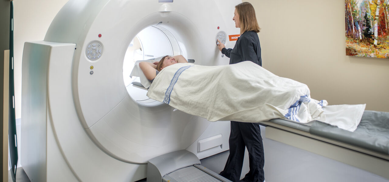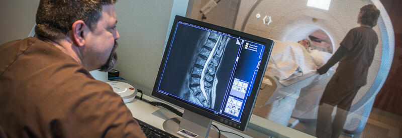

Magnetic Resonance Imaging (MRI)

Magnetic resonance imaging (MRI) is one of the safest, most comfortable imaging techniques available. MRI uses a magnetic field instead of radiation to obtain images of the body. The procedure is relatively painless and has no side-effects. MRI does not use radiation or x-rays.
The radiofrequency energy works with hydrogen, the most abundant atom in the body, which emits detectable signals in response to the energy. These signals are collected and turned into a digital image, each showing a thin slice of the body.
MRI can produce hundreds of images of the body in any plane. Due to the high degree of accuracy and detail, MRI can be a crucial imaging study in the diagnosis of multiple conditions such as: neurology, musculoskeletal and oncology disease diagnosis.
Uses for MRI
MRIs can examine the brain, spine, breasts, prostate, blood vessels (MR Angiogram), abdomen, shoulders, hips, knees and more. The procedure helps diagnose infection, cancer, stroke, neurological disorders, orthopedic/spine disorders, bone fractures, infections and torn ligaments or muscles.
 Accredited by the American College of Radiology
The MRI units at Mary Lanning Healthcare are accredited by the American College of Radiology. They are staffed by ARRT board certified MRI Technologists.
Preparation
Please wear comfortable clothing without metal fasteners, including metal buttons, zippers, eyelets and bra hooks or underwires. Do not wear hairpins, hairclips, excessive jewelry or eye makeup.
Contact your physician if you feel you need medication for pain management, anxiety or claustrophobia.
You may eat or drink normally unless otherwise instructed by your physician.
Who should avoid MRI
Patients with a pacemaker, brain aneurysm clips, certain heart valve replacements, certain neurostimulator implants and cochlear implants should not have an MRI.
After your MRI
Your MRI images are interpreted by a Board Certified Radiologist, and the results are sent to your physician’s office.


